Comparison of deconvolution techniques
Motivation
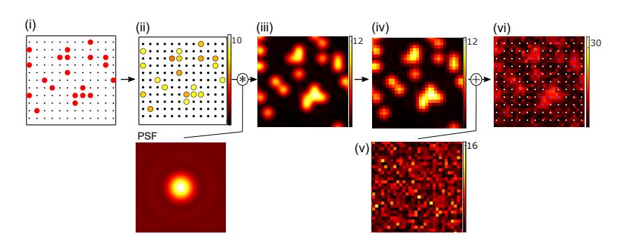
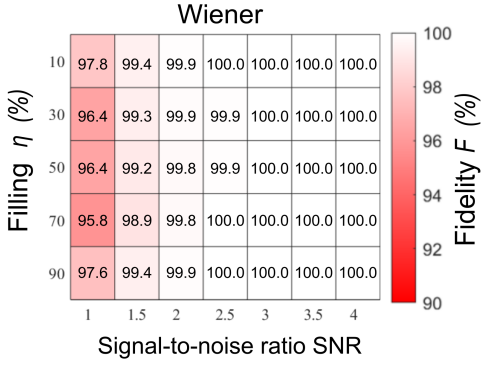
To understand the limits of single-atom imaging compare the effectiveness of three different deconvolution techniques that are commonly used to process the fluorescence images. We use simulated images to determine the fidelity of detecting single atoms in an optical lattice in the presence of noise, inhomogeneous fluorescence, for parameter regimes relevant to other quantum-gas microscope experiments.
Example for growing excitation modes
A wave packet, which is initially centred at zero momentum, quickly spreads over the complete Brillouin zone due to interactions and periodic driving. Those instabilities make quantum simulation experiments challenging.
Hyperlink
New J. Phys. 25, 083036 (2023).
Experimental setup

Experimental setup
Abstract
Quantum-gas microscopes are used to study ultracold atoms in optical lattices at the singleparticle level. In these systems atoms are localised on lattice sites with separations close to or below the diffraction limit. To determine the lattice occupation with high fidelity, a deconvolution of the images is often required. We compare three different techniques, a local iterative deconvolution algorithm, Wiener deconvolution and the Lucy-Richardson algorithm, using simulated microscope images. We investigate how the reconstruction fidelity scales with varying signal-to-noise ratio, lattice filling fraction, varying fluorescence levels per atom, and imaging resolution. The results of this study identify the limits of singe-atom detection and provide quantitative fidelities which are applicable for different atomic species and quantum-gas microscope setups.